Non-Invasive Radiosurgery: What Patients Should Know Before Treatment
Non-invasive radiosurgery, more formally called stereotactic radiosurgery, treats selected tumors and other abnormalities with highly focused beams of radiation. Despite its name, it does not involve an incision, removal of tissue, or traditional operating room surgery. Instead, sophisticated imaging and computer-guided planning direct radiation toward a defined target while limiting exposure to nearby healthy structures.
The treatment is most commonly used for conditions in the brain, including certain tumors, brain metastases, arteriovenous malformations, and trigeminal neuralgia. Similar methods are used elsewhere in the body and are often called stereotactic body radiation therapy, or SBRT. A careful evaluation is needed because radiosurgery is not appropriate for every diagnosis, size of tumor, or location.
How Focused Radiation Treats a Specific Target
Radiation damages the genetic material inside cells, making it harder for them to divide and survive. Cancer cells are often less able than healthy cells to repair this damage. Radiosurgery concentrates many small radiation beams on one target. Each individual beam passes through normal tissue at a relatively low dose, but the beams meet at the treatment area to deliver a stronger, carefully planned dose.
The word “stereotactic” refers to the use of three-dimensional coordinates. CT scans, MRI scans, and sometimes PET scans help the treatment team map the target in relation to the rest of the body. This detailed map lets the team account for structures that need protection, such as the optic nerves, brainstem, spinal cord, bowel, or lungs.
Different machines can deliver radiosurgery, including Gamma Knife systems, linear accelerators, and robotic systems. Gamma Knife is used for targets in the head and delivers many gamma-ray beams from fixed positions. Linear accelerator systems can treat the brain and body, often rotating around the patient to deliver radiation from multiple angles. The best platform depends on the condition, target location, size, and the expertise available at the treatment center.
Radiosurgery does not usually make a tumor disappear immediately. The treatment causes changes within the targeted cells over time. Some tumors shrink gradually, some stop growing, and vascular abnormalities may close off over months or years. Follow-up imaging is therefore part of treatment, not merely an administrative step after the procedure. For targets that move with breathing or other natural body motion, robotic systems can add real-time tracking that adjusts the radiation beam as the target changes position. Patients considering this type of treatment can learn more at https://drjameswilson.co.uk/treatments/cyberknife/.
Conditions and Patient Factors That Shape Eligibility
For brain treatment, radiosurgery may be considered for small or limited numbers of metastatic tumors, certain benign tumors such as vestibular schwannomas, pituitary tumors, and meningiomas, or blood-vessel abnormalities known as arteriovenous malformations. It can also be used to treat trigeminal neuralgia, a condition that causes severe facial pain. Its purpose differs by diagnosis, ranging from controlling tumor growth to relieving symptoms or closing abnormal blood vessels.
Target size matters because focused radiation has limits. A larger lesion may expose too much surrounding tissue to radiation if treated in one session. In those cases, clinicians may recommend surgery, conventional radiation given over several weeks, or stereotactic treatment divided into several sessions. Dividing treatment is called fractionation, and it can allow normal tissue more time to recover between doses.
Location is equally significant. A small tumor in an area away from sensitive structures may be a good candidate for a single high-dose treatment. A similarly sized target next to the optic nerves or brainstem requires a more cautious plan because even a small amount of swelling or radiation injury could affect vision, movement, speech, swallowing, or other functions.
The decision also reflects the person’s broader health picture. Doctors consider prior radiation exposure, surgery history, medication use, symptoms, ability to lie still, and the goals of care. Radiosurgery can be used alone, after surgery, alongside drug treatment, or as part of a longer plan that includes monitoring. A recommendation should be tailored to the diagnosis rather than based solely on the appeal of avoiding an operation.
What Planning and Treatment Day Usually Involve
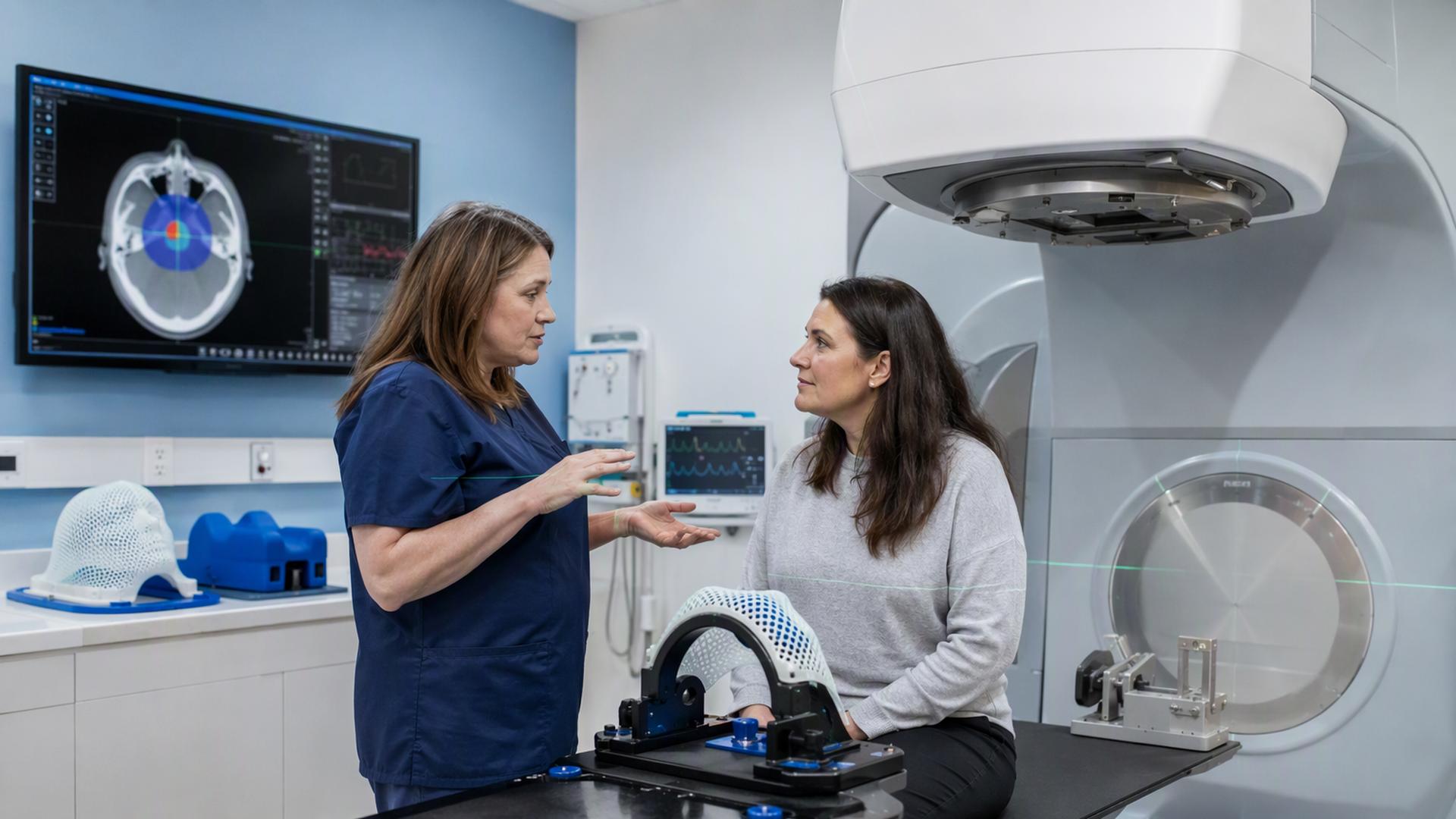
Planning begins with imaging that shows the target as accurately as possible. For brain radiosurgery, patients may have a lightweight frame secured to the head or a custom-fitted mask that helps prevent movement. Body treatments use immobilization devices, positioning supports, and sometimes breathing-management techniques. These measures may sound restrictive, but their purpose is to make radiation delivery precise.
A radiation oncologist, medical physicist, dosimetrist, and other specialists develop the treatment plan. They outline the target and nearby normal tissues on imaging scans, then calculate beam angles, dose, and the boundaries of the treatment area. This planning process can take longer than the actual radiation session because accuracy is central to both effectiveness and safety.
Preparation instructions vary. A care team may ask a patient to avoid food or drink for a period before treatment if sedation is planned, or to adjust medications such as blood thinners, diabetes drugs, or anti-seizure medicines. Patients should provide a complete medication list, report allergies and implanted devices, and ask which regular prescriptions should be taken on treatment day. A support person may be needed if sedation is used.
During treatment, patients do not feel the radiation itself. The machine may move around the treatment table, and staff monitor the session from a nearby control room while communicating through cameras and an intercom. A single-session procedure can last from minutes to several hours, depending on the system and complexity of the plan. Most people go home the same day, although recovery arrangements should be confirmed in advance.
Side Effects, Follow-Up, and When to Seek Help
Side effects depend on the body area treated, radiation dose, and nearby organs. Fatigue is common after radiation treatment and can last for days or weeks. For brain radiosurgery, short-term effects can include headache, nausea, scalp irritation, or temporary swelling near the treated site. Doctors may prescribe corticosteroids to reduce swelling when appropriate.
Some effects appear later rather than immediately. Radiation can cause inflammation or tissue changes at the treatment site months after brain radiosurgery. In uncommon cases, radiation necrosis can occur, meaning treated tissue becomes damaged and may cause swelling or symptoms that resemble tumor growth. Follow-up scans help clinicians distinguish treatment-related changes from progression of the original condition.
Risks vary with the target’s location. Treatment near nerves can affect hearing, vision, balance, sensation, or facial movement. SBRT to the chest, abdomen, or pelvis can irritate nearby organs, potentially causing symptoms such as cough, swallowing discomfort, bowel changes, urinary issues, or pain. The treatment team should explain the risks most relevant to the individual plan rather than offering only a general list.
Contact the care team promptly for new or worsening neurologic symptoms, severe headache, persistent vomiting, confusion, seizures, sudden weakness, breathing difficulty, fever, or unusual pain. Scheduled MRI, CT, or other imaging appointments should also be kept even when a person feels well. Radiosurgery outcomes are often judged over time, and monitoring allows clinicians to respond if swelling, recurrence, or another treatment-related issue develops.
Preparing for an Informed Treatment Decision
Non-invasive radiosurgery can offer a precise treatment option with no surgical incision and little interruption to daily life, but it remains a serious medical procedure with benefits, limits, and potential side effects. Patients can prepare by asking about the treatment goal, alternatives, expected timeline of results, likely short-term effects, long-term monitoring, and who to contact after treatment. Clear answers from a multidisciplinary care team help turn a complex decision into a plan that fits both the medical condition and the patient’s priorities.